Body imaging provides a unique window into the human body, offering detailed images of organs, tissues, and bones. These technologies enable health professionals to observe the body’s internal structures without the need for invasive procedures. Understanding how body imaging works can help you feel more prepared and informed about your health. It is a powerful tool for observing the intricate workings of your internal systems.
What Is Body Imaging?
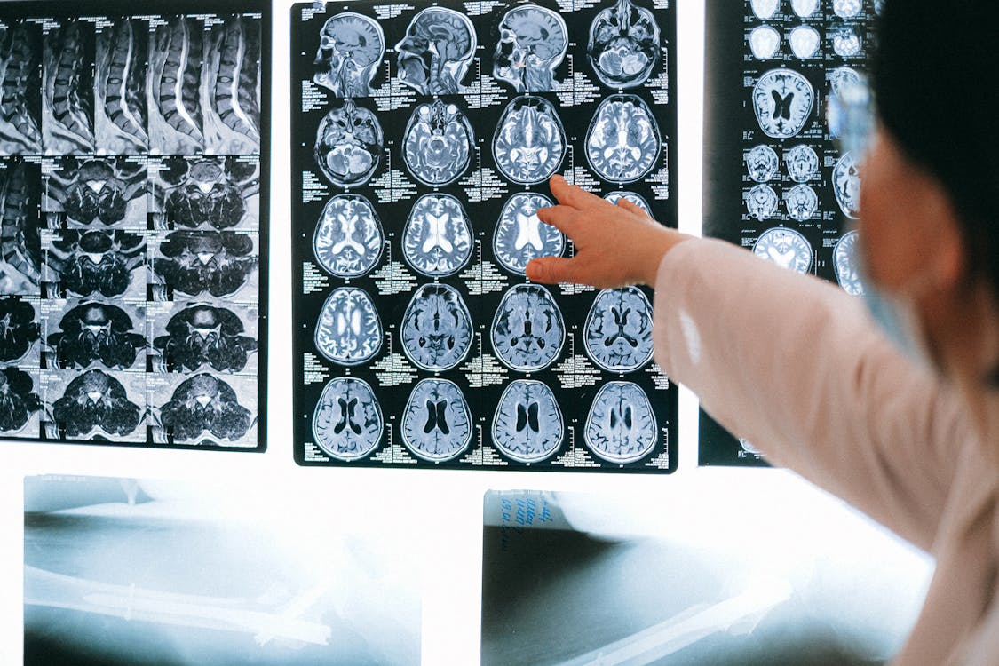
Body imaging refers to a group of non-invasive techniques used to create images of the body’s interior. These methods utilize different forms of energy, such as X-rays, sound waves, or magnetic fields, to generate detailed visuals. Common types of body imaging include computed tomography (CT) scans, magnetic resonance imaging (MRI), and ultrasound. Each modality provides a distinct view of tissues and structures.
These scans give health professionals a clear picture of your organs and their condition. The images can show the size, shape, and texture of internal body parts. This information helps in observing the body over time and tracking any developments. The process is straightforward and provides a wealth of information.
How Do Scans Detect Early Changes?
Scans detect early changes by creating high-resolution images that can reveal minute abnormalities. These technologies are sensitive enough to spot subtle differences in tissue density, size, or shape that might otherwise go unnoticed. An MRI, for instance, uses a strong magnetic field and radio waves to generate detailed images of soft tissues, making it helpful in examining the brain, muscles, and abdominal organs. A CT scan combines a series of X-ray images from different angles to create cross-sectional pictures.
These detailed images allow for a comparative analysis over time. A radiologist can compare a current scan to a previous one to identify any new or evolving changes. This comparison helps in monitoring organ health and detecting shifts from a baseline state. The ability to visualize these small variations is a key aspect of modern diagnostic approaches.
What Conditions Can Imaging Reveal?
Body imaging can reveal a wide range of conditions by showing structural changes in organs. These scans help provide a visual basis for understanding what is happening inside your body. The detailed pictures can highlight areas that may require further attention.
Different imaging techniques are suited for identifying various conditions. Some of the conditions that imaging can help visualize include:
- Liver conditions: Ultrasound, CT, and MRI scans show changes in the liver’s size and texture. They can also detect fatty deposits or other structural differences.
- Kidney issues: Imaging can reveal blockages, cysts, or changes in kidney size. These scans provide clear pictures of the urinary tract.
- Heart-related changes: Cardiac MRI and CT angiography provide detailed images of the heart’s structure and blood vessels. They can visualize the heart muscle and its function.
- Gallbladder problems: An ultrasound is often used to look for gallstones or inflammation in the gallbladder. It provides a real-time view of the organ.
These are just a few examples of how imaging can provide insight into your organ health. The information gathered from these scans supports a more complete picture of your overall well-being.
Consult a Specialist
The results of any body imaging scan are complex and require a radiologist, a medical doctor trained in reading these images, to analyze the findings and prepare a detailed report. Your healthcare provider will then review this report with you and explain what the results mean for your health. If you have any questions or concerns, discussing them with your provider is the best next step, as they can determine if body imaging is appropriate and guide you through the process. Open communication with your specialist is key to navigating your health journey effectively.