Medical imaging provides non-invasive methods to view internal structures. Magnetic Resonance Imaging (MRI) and ultrasound are two common modalities, each using different principles to create images. The debate of ultrasound vs mri often depends on the specific clinical question. Each technology has distinct strengths and limitations that influence diagnostic accuracy for different conditions. The differences between the technologies help explain why one imaging method is sometimes preferred.
Capabilities and Scope
What are the main differences in an ultrasound vs MRI? Mainly the difference lies in their imaging scope and the types of tissues they can visualize.
Ultrasound Capabilities
An ultrasound provides high-resolution images of soft tissues, particularly those close to the skin’s surface. It shows muscles and organs in real time, which enables a doctor to assess dynamic motion in the tissues. This capability may allow for identification of tendon injuries or blood clots and for precision guidance in injections.
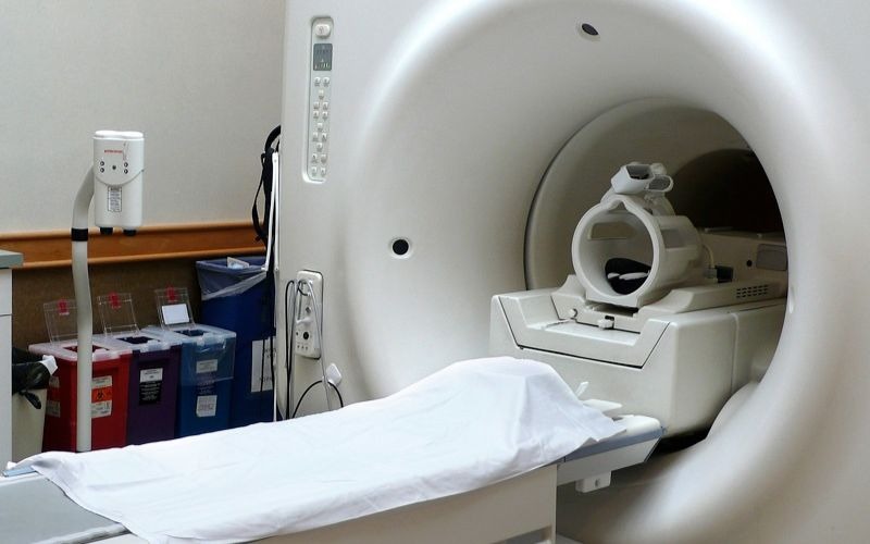
MRI Capabilities
An MRI offers a broader and deeper view, producing 3D images of entire body regions. While an ultrasound visualizes the soft tissues around a joint, an MRI can penetrate to show internal joint structures, including cartilage and bones. This capability may be helpful for diagnosing conditions that affect deep or large areas, like spinal cord injuries or complex damage within joints. An MRI delivers a more complete picture when a wider field of view is required.
Technology and Applications
Ultrasound technology uses high-frequency sound waves. A transducer sends and receives these waves, creating echoes that bounce back from tissues and organs. The images help visualize soft tissue, fluid-filled spaces, and blood flow. Common uses include monitoring fetal development, assessing heart function, and imaging the liver, gallbladder, and kidneys. Because it uses no radiation, ultrasound is often preferred for pregnant patients.
MRI uses a strong magnetic and radio waves to generate detailed cross-sectional images of soft tissue, such as the brain, spinal cord, joints, and internal organs. Providers may use an MRI to examine bodily areas with a high degree of anatomical detail. A contrast agent may also be used to enhance imaging.
Accuracy and Limitations
An ultrasound’s diagnostic accuracy depends on the operator’s skill. Image quality can be influenced by patient size or abdominal gas. Ultrasound is highly accurate for gallstones or for guiding biopsies. Its ability to provide dynamic imaging allows clinicians to observe organ function and blood flow, which is a unique advantage in certain diagnostic scenarios.
MRI’s soft tissue contrast can help achieve high diagnostic accuracy, making it beneficial for diagnosing brain, spine, and complex joint issues. The detail provided by MRI can help identify subtle tissue abnormalities and differentiate between various tissue types. Scans may take longer, and not all patients can receive an MRI because of metal implants.
Ultrasound vs MRI: Learn More
Deciding between an ultrasound and an MRI depends on the clinical setting, body part, and the suspected condition. Both methods offer valuable diagnostic insights to the provider and patient. Consult a healthcare provider near you to learn more about your options for ultrasounds and MRIs.